
Spine pain
Degenerative Disc Disease
Degenerative disc disease is disc aging, not a disease. When it actually causes pain, disc vs facet pain, and treatment options in Houston.
Degenerative disc disease is a misleading name for a normal process: spinal discs dry out, flatten and crack with age, and by 60 nearly everyone has it on an MRI. It becomes a problem only when a worn disc is the source of pain, which is true for a minority of people who have the finding. Gulf Coast Pain & Spine works out whether your pain is coming from the disc, the facet joints behind it, or a nerve, and treats that source rather than the MRI report, at its Houston and Webster offices and, from November 2026, in Pearland.
Key facts
| What it is | Age-related loss of water, height and structure in the discs between the vertebrae, most often at L4–5 and L5–S1 in the low back and C5–6 and C6–7 in the neck. It is a description of imaging, not a diagnosis of pain. |
|---|---|
| How common | Disc degeneration is present on MRI in about 37% of pain-free 20-year-olds and 96% of pain-free 80-year-olds. Genetics accounts for most of the variation; smoking, heavy loading and obesity add to it. |
| When it causes pain | When the outer disc wall is torn and inflamed (discogenic pain), when the flattened disc overloads the facet joints behind it, when it narrows a nerve's exit, or when the adjacent bone becomes inflamed (Modic changes, also called vertebrogenic pain). |
| See a specialist when | Back or neck pain has lasted more than 6–12 weeks, flares repeatedly, is worse sitting and bending, or comes with arm or leg symptoms. |
| Treatments we offer | Coordinated therapy and medication review, epidural steroid injections for nerve involvement, medial branch blocks and radiofrequency ablation for the facet component, basivertebral nerve ablation for confirmed vertebrogenic pain, and, with an explicit evidence and cost discussion, intradiscal options. |
When to get emergency care
Disc degeneration itself is not dangerous. Seek emergency care or call 911 if your back or neck pain comes with:
- Numbness in the groin or inner thighs, trouble urinating, or loss of bowel or bladder control.
- Weakness in an arm or leg that is new or getting worse, a dragging foot, hand clumsiness, or a change in balance or walking.
- Fever or chills with spine pain, especially with diabetes, a recent infection or a recent spine procedure; discs and adjacent bone can become infected (discitis).
- Pain after a fall or accident, particularly over age 65 or with osteoporosis, until a fracture is ruled out.
- A history of cancer with new spine pain, unexplained weight loss, or pain that is worst at night and does not ease with rest.
Not a disease: what disc degeneration actually is
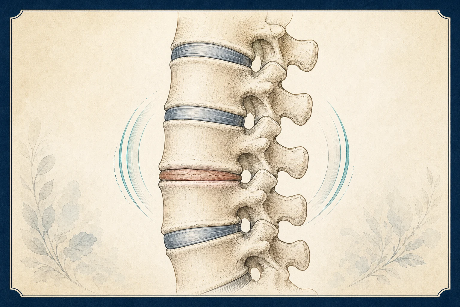
A healthy disc is about 80% water. It has a gel center that absorbs load and a fibrous outer ring that holds it in place. Starting in the twenties, the center gradually loses water and the disc becomes shorter, stiffer and darker on MRI (a "black disc"). Small cracks (fissures) form in the outer ring. Bone spurs develop at the edges of the vertebrae, and the bone next to the disc can show inflammation or fatty change (Modic changes). Radiologists call the whole picture degenerative disc disease or spondylosis.
None of this is a disease. It is the spine's version of gray hair, and it happens to everyone at a pace set mostly by genetics. In a pooled analysis of more than 3,000 people with no back pain at all, disc degeneration was present in 37% of 20-year-olds, 68% of 40-year-olds, 88% of 60-year-olds and 96% of 80-year-olds. Disc height loss, bulges and Modic changes were similarly common. If your MRI report lists degenerative disc disease, it is describing your age more than your pain.
That said, a worn disc can hurt, and for some people it is the main source of chronic low back or neck pain. The work of the first visit is to decide whether your pain is one of those cases, and if so, whether it is the disc itself, the joints behind it, or the nerve next to it that needs treatment.
How a degenerated disc produces pain
A disc can generate pain through four routes, and each has its own treatment path.
Discogenic pain. The outer third of the disc wall has nerve endings. When a fissure reaches that zone and becomes inflamed, the disc itself hurts. Pain is central, deep, and worse with sitting, bending forward, lifting and coughing, all of which raise pressure inside the disc. It eases with standing, walking and lying down. Flares last days to weeks and recur.
Vertebrogenic pain. When the endplate, the thin layer of bone between the disc and the vertebra, is damaged, the bone becomes inflamed. This shows as Modic type 1 (inflammatory) or type 2 (fatty) changes on MRI, and the pain is carried by a specific nerve inside the vertebra, the basivertebral nerve. This is a distinct, treatable subtype.
Facet joint overload. As the disc flattens, more load shifts to the two facet joints behind it. Facet pain is off to the side, worse arching backward and twisting, stiff in the morning, and better sitting, which is the opposite of discogenic pain. Many people have both at the same level. See facet joint syndrome.
Nerve involvement. A flattened, bulging disc and its bone spurs can narrow the opening where a nerve root exits (foraminal stenosis) or herniate into it, producing arm or leg pain along that nerve. See sciatica, cervical radiculopathy, herniated disc and spinal stenosis.
Symptoms: discogenic versus facet pain
The pattern of your pain does more to identify the source than the MRI does.
- Discogenic: central low back or neck pain; worse sitting, bending forward, driving, lifting, coughing; better standing and walking; flares that last days to weeks; pain sometimes spreads to the buttocks or shoulders but not in a nerve pattern.
- Facet: pain to one or both sides of the spine; worse arching, twisting, standing for long periods; stiff in the morning; better sitting and bending forward; tender directly over the joints.
- Vertebrogenic: behaves like discogenic pain, central and worse sitting, often in people whose MRI shows Modic changes at one or two levels and whose pain has lasted more than 6 months.
- Nerve: pain that travels down an arm or leg past the elbow or knee with tingling, numbness or weakness.
Discogenic and facet pain frequently coexist and the pattern can be mixed. That is one reason a treatment aimed at only one of them sometimes helps only partly, and why we use diagnostic blocks to sort the components rather than guess.
How we diagnose it
The exam tests which movements reproduce the pain: flexion and sitting load the disc, extension and rotation load the facets. We check strength, reflexes and sensation in the limbs for nerve involvement, press over the facet joints, and examine the SI joints and hips, which imitate low back pain.
Imaging. An MRI is ordered when pain has persisted more than 6 weeks, when there are nerve symptoms or red flags, or to plan a procedure. We read it level by level for the things that change management: a fissure or herniation at the level that matches your symptoms, Modic changes at the endplates, foraminal narrowing, and facet arthritis. We do not treat degeneration that is at a level or side that does not match your pain. Plain X-rays with bending views are used when instability (spondylolisthesis) is suspected.
Diagnostic blocks. If the pattern suggests a facet component, medial branch blocks on two separate days test it: 80% or more relief confirms the facets and opens the door to ablation. If it suggests the nerve, a transforaminal injection or selective nerve root block at the suspected level is both a test and a treatment. Discogenic pain is diagnosed by the pattern and the MRI; provocative discography, which injects the disc to see if it reproduces pain, is now used rarely and mainly before surgery, because it can accelerate degeneration of the tested disc.
Treatment options, in order
Because disc degeneration is permanent and usually painless, treatment is aimed at the pain, not the picture. The goal is a spine that tolerates your life, not an MRI that looks younger.
- Exercise, weight and load management, medication review
The strongest long-term evidence for chronic disc-related back pain is regular exercise: walking, core and hip strengthening, and graded return to activity. NSAIDs for flares if safe for you; duloxetine for chronic pain in some people. Opioids are not recommended for chronic back pain. Stop smoking; it accelerates degeneration and worsens outcomes of every treatment. Learn more →
- Physical therapy
A program built around your pattern: extension-biased for discogenic pain, flexion-biased for facet pain, plus hip and core strength for both. Typically 6–12 weeks, with a home program that continues. Learn more →
- Epidural steroid injection
When a degenerated disc is irritating a nerve root and producing arm or leg pain. Relief typically begins within 2–7 days and lasts weeks to months. The evidence is good for limb pain and weak for back pain alone, so we do not use epidurals for purely discogenic pain. Learn more →
- Medial branch blocks, then radiofrequency ablation
For the facet component, which is confirmed by two blocks. Ablation gives typically 6–12 months or longer of relief and can be repeated. Learn more →
- Basivertebral nerve ablation (for vertebrogenic pain)
For chronic low back pain of more than 6 months with Modic type 1 or 2 changes at L3 to S1 on MRI that has not responded to 6 months of conservative care. A probe placed through the pedicle heats the basivertebral nerve inside the vertebra. Two randomized trials showed durable improvement in pain and function, and Medicare covers it for patients who meet those criteria; commercial coverage varies. Learn more →
- Intradiscal options, including VIA Disc
VIA Disc is an injection of processed donor disc tissue (an allograft) into the degenerated disc. The evidence is limited to one industry-sponsored randomized trial with short follow-up; it is regulated as a human tissue product rather than approved as a drug or device for disc pain, and it is typically not covered by insurance, so patients pay out of pocket. We discuss it only after the steps above, with that caveat stated plainly. Read the full evidence and cost statement before considering it. Learn more →
- Surgical referral
Fusion or disc replacement for single-level discogenic pain is an option of last resort with mixed evidence: trials comparing fusion to intensive rehabilitation show similar results at two years. We refer when there is instability, progressive nerve compression, or disabling pain after every appropriate step above. Learn more →
What you can do now
- Walk every day. Discs get their nutrition from movement; there is no medication that does what a daily walk does for a degenerated disc.
- Break up sitting. Stand or walk for two minutes every 30 minutes. Sitting loads the disc more than standing does.
- Lift with your hips, close to the body, and do not twist under load.
- Lose weight if you carry extra; every pound of body weight adds several pounds of load on the lumbar discs when bending.
- Stop smoking. Nicotine narrows the small vessels that feed the disc and roughly doubles the rate of degeneration and the risk of persistent pain.
- Do not chase the MRI. Repeat imaging for the same pain rarely changes anything and often raises worry. Bring the MRI you have; it is enough for most decisions.
- Track your flares: what triggered them, how long they lasted, what settled them. Sitting and bending triggers point to the disc; arching and twisting point to the facets.
Frequently asked questions
Is degenerative disc disease actually a disease?
No. It is the normal aging of the disc and is present on MRI in most adults by their forties and nearly all by their eighties, including people with no pain. The name is a leftover from older radiology terminology. It matters only when a worn disc is the source of your pain, which we determine from the pattern and exam, not the report.
Will degenerative disc disease get worse?
The disc will continue to age, as everyone's does, but pain does not track with the imaging. Many people with worsening MRI findings have less pain over time as the disc stiffens and settles. Flares tend to become less frequent with consistent exercise and weight control. Progression to nerve compression or instability is uncommon and is what we monitor for.
How do I know if my pain is from the disc or the facet joints?
Disc pain is central, worse sitting and bending forward, and eases standing. Facet pain is to the side, worse arching backward and twisting, stiff in the morning, and eases sitting. Many people have both. Medial branch blocks test the facet component directly; the disc component is judged by the pattern and the MRI.
What are Modic changes?
Inflammatory (type 1) or fatty (type 2) changes in the bone next to a degenerated disc, visible on MRI. They indicate endplate damage and identify a subgroup whose pain comes from the vertebra itself (vertebrogenic pain). That subgroup can be treated with basivertebral nerve ablation, which has randomized-trial evidence and Medicare coverage for patients who meet the criteria.
Does the VIA Disc procedure work?
The evidence is thin. VIA Disc injects processed donor disc tissue into a degenerated disc. There is one industry-sponsored randomized trial with short follow-up, it is not FDA-approved as a drug or device for disc pain, and insurance generally does not cover it, so it is a cash-pay procedure. We present it only after conservative care, facet and nerve treatments have been exhausted and with those limits stated. Our VIA Disc page sets out the full evidence and cost picture.
Can degenerative disc disease cause leg or arm pain?
Indirectly. A flattened disc and its bone spurs can narrow the opening where a nerve leaves the spine, or the disc can herniate into it, producing sciatica or cervical radiculopathy. That nerve pain is treated as a nerve problem, usually with a targeted epidural injection, and it often improves on its own over weeks.
Should I have a spinal fusion for degenerative disc disease?
Rarely. Trials comparing fusion with intensive rehabilitation for chronic discogenic back pain show similar outcomes at two years, and fusion changes load on the adjacent levels. Surgery is reserved for instability, progressive nerve compression, or disabling pain after every nonsurgical step has been tried. Most people with DDD never need it.
Sources
- Systematic literature review of imaging features of spinal degeneration in asymptomatic populations (Brinjikji et al.) (opens in new tab) — American Journal of Neuroradiology, 2015
- Low Back Pain Fact Sheet (opens in new tab) — National Institute of Neurological Disorders and Stroke (NIH)
- Noninvasive Treatments for Acute, Subacute, and Chronic Low Back Pain: A Clinical Practice Guideline (2017) (opens in new tab) — American College of Physicians
- Low Back Pain (opens in new tab) — American Academy of Orthopaedic Surgeons, OrthoInfo
- Comprehensive Evidence-Based Guidelines for Facet Joint Interventions in the Management of Chronic Spinal Pain (2020) (opens in new tab) — American Society of Interventional Pain Physicians
Request an appointment
Call (832) 916-2075 or request an appointment online. New patients are welcome; the team confirms insurance and referral requirements before scheduling.