
Spine pain
Herniated Disc (Bulging, Protruded, Extruded or Sequestered Disc)
Bulging, protruded, extruded, sequestered: what your MRI words mean, why most herniated discs shrink on their own, red flags, and treatment in Houston.
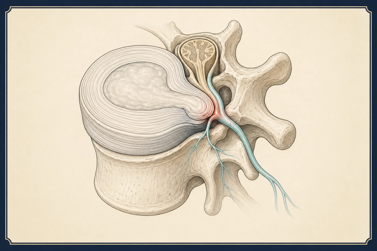
A herniated disc is a spinal disc whose soft center has pushed through a tear in its outer wall. It matters only when it presses on or inflames a nerve, and even then most herniations shrink on their own over months; the larger the herniation, the more likely it is to resorb. "Bulging," "protruded," "extruded" and "sequestered" are points on one spectrum, not separate diseases. Gulf Coast Pain & Spine matches the MRI to your symptoms, treats nerve pain with targeted epidural injections while the disc heals, and refers for surgery only when weakness or unrelenting pain requires it, at its Houston and Webster offices and, from November 2026, in Pearland.
Key facts
| What it is | Displacement of disc material beyond its normal boundary. In the low back, most occur at L4–5 and L5–S1; in the neck, at C5–6 and C6–7. Thoracic herniations are rare. |
|---|---|
| Most common causes | Age-related drying and weakening of the disc wall, then a trigger: lifting with a twist, a fall, prolonged sitting or driving, or nothing identifiable at all. Smoking and heavy physical work raise the risk. |
| Typical course | Nerve pain from a herniation improves in about 75% of people within 6–12 weeks. On repeat MRI, roughly two-thirds of extrusions and nearly all sequestered fragments shrink or disappear within a year. |
| See a specialist when | Arm or leg pain has lasted more than 4–6 weeks, there is any weakness, or pain is severe enough to stop work or sleep. |
| Treatments we offer | Epidural steroid injections (lumbar, cervical, caudal), selective nerve root blocks, coordinated therapy and medication, and surgical referral when criteria are met. |
When to get emergency care
Go to an emergency room or call 911 if you have a known or suspected disc herniation with any of these:
- Numbness in the groin, inner thighs or around the rectum, difficulty starting or stopping urine, or loss of bowel or bladder control. A large central lumbar herniation can compress the cauda equina; outcomes depend on surgery within 24–48 hours.
- A foot you cannot lift (foot drop), weakness that is getting worse over hours or days, or weakness in both legs.
- For a neck herniation: clumsy hands, trouble with buttons or handwriting, or unsteady walking. These mean the spinal cord is compressed (myelopathy).
- Symptoms in both arms or both legs at the same time.
- Fever or chills with back or neck pain, or a history of cancer with new spine pain and unexplained weight loss.
- Severe pain after a fall or accident until a fracture is excluded.
Mild, stable weakness, such as slightly less push-off in the calf, is common and is followed closely rather than operated on. Weakness that is progressing is the difference.
What a herniated disc is, and what the MRI words mean
Each disc has a tough outer ring (annulus) and a gel-like center (nucleus). With age the nucleus dries and the annulus develops small tears. A herniation is nucleus material pushing through those tears beyond the edge of the vertebra. Radiologists describe how far it has gone using standard terms, and patients are often frightened by words that describe the same process at different stages:
- Bulging disc. The whole disc edge extends slightly beyond the vertebra, all the way around or across more than a quarter of its circumference. This is not a herniation. It is a normal finding in most adults over 40 and is rarely the cause of pain by itself.
- Protrusion. A focal herniation where the piece pushing out is still wider at its base than at its tip. Contained by the outer fibers of the annulus.
- Extrusion. The herniated piece has pushed through the annulus and is now wider than the opening it came through, like toothpaste out of a tube. Often called a "ruptured" or "slipped" disc.
- Sequestration. A fragment has broken free from the disc and lies loose in the spinal canal.
Here is the part that surprises people: the further along the spectrum, the more likely the herniation is to go away. The body treats extruded and sequestered disc material as foreign and absorbs it. In pooled studies of repeat MRI, about 13% of bulges, 41% of protrusions, 70% of extrusions and 96% of sequestrations showed regression. A "large extruded disc" on a report sounds worse than a bulge and usually has the better natural history.
Causes and how common disc findings are
Discs weaken with age, genetics, smoking, and years of load; the herniation itself is often triggered by bending and twisting under load, a fall, a long drive, or a sneeze. Many people cannot identify any event. The lowest two lumbar discs (L4–5 and L5–S1) take the most bending stress and account for about 90% of lumbar herniations. In the neck, C5–6 and C6–7 are the most mobile levels and the most commonly affected.
Disc findings are extremely common on MRI in people without pain. In a review of more than 3,000 asymptomatic adults, disc bulges were present in 30% of 20-year-olds and 84% of 80-year-olds, and protrusions in 29% of 20-year-olds and 43% of 80-year-olds. A herniation on your MRI is only the explanation for your pain if it is at the level and on the side that matches your symptoms. That match, not the MRI report, is what we treat.
Symptoms: lumbar versus cervical
A herniation produces symptoms in two ways. It can irritate the outer wall of the disc, causing pain in the back or neck itself (discogenic pain), and it can compress or inflame the adjacent nerve root, causing pain along that nerve into a limb (radiculopathy). Most people with a symptomatic herniation have more limb pain than spine pain, and the limb pain is what the treatment targets.
Lumbar herniation. Low back pain followed within days by shooting pain down the buttock and one leg, usually below the knee, with tingling or numbness in the foot: sciatica. Worse with sitting, bending forward, coughing and sneezing; often better standing or lying down. An L4–5 herniation typically compresses the L5 root (outer calf, top of the foot, big toe; weak toe and ankle lift). An L5–S1 herniation compresses S1 (back of the calf, outer foot, small toes; weak push-off; reduced ankle reflex). A large central herniation can affect both legs and the bladder, which is the cauda equina emergency described above.
Cervical herniation. Neck pain with pain shooting across the shoulder and down one arm to specific fingers, tingling or numbness in those fingers, and sometimes weakness: cervical radiculopathy. Worse with looking up or turning toward the painful side; often eased by resting the hand on top of the head. C5–6 affects C6 (thumb and index finger, biceps); C6–7 affects C7 (middle finger, triceps). A central cervical herniation can press on the spinal cord itself and cause hand clumsiness and gait change, which is myelopathy and needs prompt surgical evaluation.
Look-alikes. SI joint pain and hip arthritis mimic lumbar herniation but do not go below the knee and do not cause true numbness. Carpal tunnel syndrome mimics a C6 herniation but wakes people at night and spares the forearm. Peripheral neuropathy affects both feet in a stocking pattern.
How we diagnose it
The history and exam usually identify the level before any imaging. For the low back we map where the pain and numbness go, test strength in the hip, knee, ankle and toes, check knee and ankle reflexes, and perform the straight-leg raise; leg pain reproduced between 30 and 70 degrees is the classic sign of nerve root tension. For the neck we map the fingers involved, test each muscle group and reflex, and perform Spurling's test. In both regions we examine the joints and peripheral nerves that imitate a herniation.
MRI is the test that shows the disc and the nerve, and it is ordered when limb pain has lasted more than 4–6 weeks, when there is weakness, when a red flag is present, or before an injection. It is not ordered on day one for typical improving sciatica, because the result rarely changes early management and often shows unrelated findings. CT myelography is used when MRI is not possible. EMG is used when the exam and MRI disagree or to separate a root problem from a peripheral nerve problem. When MRI shows possible compression at two adjacent levels, a selective nerve root block identifies the level responsible; this is a test, not a treatment.
Treatment options, in order
Because most herniations resorb and most nerve pain settles within 6–12 weeks, the strategy is to control pain and protect the nerve while that happens. Injections shorten the painful window; surgery is for the minority whose weakness or pain does not allow waiting.
- Weeks 1–6: activity and medication
Stay active within pain limits; avoid bed rest beyond a day or two, heavy lifting and deep forward bending. NSAIDs for 1–2 weeks if safe for you; a short oral steroid taper for severe early nerve pain; a muscle relaxant for sleep if needed. Opioids only briefly, if at all. Learn more →
- Physical therapy
Directional exercises that draw pain out of the limb toward the spine, nerve gliding, walking progression, and later core and hip strengthening. Usually started in weeks 2–3. Learn more →
- Epidural steroid injection
Steroid delivered under X-ray guidance to the inflamed nerve root: transforaminal or interlaminar in the low back, interlaminar at C7–T1 in the neck under specific safety protocols. Relief typically begins within 2–7 days and lasts weeks to months; the evidence is clearest for limb pain and weaker for back pain alone. Limited to 3–4 per year. Learn more →
- Caudal epidural injection
An alternative route through the tailbone for lower lumbar herniations, multilevel disease, or after previous surgery. Learn more →
- Surgical referral
Microdiscectomy in the low back, or discectomy with fusion or disc replacement in the neck. Referred for progressive or significant weakness, cauda equina or myelopathy signs, or disabling limb pain after 6–12 weeks of full nonsurgical care with a matching MRI. In the SPORT trial, surgery relieved leg pain faster, and both groups improved substantially by two years. Learn more →
- Neuromodulation
Spinal cord or dorsal root ganglion stimulation for limb nerve pain that persists after surgery. Learn more →
What you can do now
- Keep walking. Short walks several times a day. Sitting raises disc pressure more than standing or lying, so limit sitting to 20–30 minutes at a time in the first weeks.
- Hinge, do not bend. Bend at the hips and knees to pick things up; avoid twisting while lifting.
- Find a relief position. Lying on the back with knees over a pillow, or on the side with a pillow between the knees, unloads a lumbar disc. For the neck, one supportive pillow and no stomach sleeping.
- Use heat on the back or neck to ease the muscle guarding around the nerve.
- Take NSAIDs on a schedule for a week or two if your doctor has said they are safe.
- Test strength weekly: heel walk and toe walk for the leg; grip and pushing against a wall for the arm. Report any loss.
- Stop smoking. Smoking impairs disc nutrition and healing and raises the risk of a repeat herniation.
Frequently asked questions
Is a bulging disc the same as a herniated disc?
No. A bulge is the whole disc edge extending slightly beyond the vertebra, a normal finding in most adults over 40 and rarely painful by itself. A herniation is a focal piece of the disc's center pushing through the outer wall. Protrusion, extrusion and sequestration are increasing degrees of herniation.
Can a herniated disc heal on its own?
Yes, in most cases. The body absorbs herniated disc material over months. About 70% of extrusions and nearly all sequestered fragments shrink on repeat MRI within a year. The annulus does not fully regain its original strength, so the same level can herniate again, which is why core strength and lifting habits matter afterward.
My MRI says extruded disc. Do I need surgery?
Not because of the word. Extruded discs are actually more likely to resorb than smaller protrusions. Surgery is decided by symptoms: progressive weakness, cauda equina or myelopathy signs, or disabling limb pain that has outlasted 6–12 weeks of complete nonsurgical treatment. Most people with an extruded disc recover without surgery.
How long does herniated disc pain last?
Limb pain usually peaks in the first 2–4 weeks and improves substantially by 6–12 weeks. Numbness and mild weakness resolve more slowly, over months, and a small residual patch of numbness is not unusual. Back pain from the disc itself can persist longer and behaves more like degenerative disc pain.
Does an epidural steroid injection shrink the disc?
No. It reduces the inflammation around the nerve so the pain drops while the disc shrinks on its own. Relief usually begins within 2–7 days and lasts weeks to months. Roughly half of people get meaningful relief from the first injection, and the benefit is strongest for limb pain rather than back pain alone.
What is the difference between a lumbar and a cervical herniation?
The process is identical; the location changes the symptoms and the risks. A lumbar herniation causes sciatica and, if very large and central, can compress the cauda equina. A cervical herniation causes arm pain and, if central, can press on the spinal cord itself, causing hand clumsiness and gait change. Cervical injections also require additional safety measures because of the cord's proximity.
Can I work out with a herniated disc?
Walking, swimming and stationary cycling are safe for almost everyone once the acute pain settles. Avoid heavy deadlifts, sit-ups, and loaded forward bending or twisting until symptoms have been gone for several weeks, then rebuild gradually. Any exercise that pushes pain further down the limb should stop.
Sources
- Herniated Disk in the Lower Back (opens in new tab) — American Academy of Orthopaedic Surgeons, OrthoInfo
- Lumbar disc nomenclature: version 2.0 (Fardon et al.) (opens in new tab) — The Spine Journal / NASS, ASSR, ASNR, 2014
- Systematic literature review of imaging features of spinal degeneration in asymptomatic populations (Brinjikji et al.) (opens in new tab) — American Journal of Neuroradiology, 2015
- The probability of spontaneous regression of lumbar herniated disc: a systematic review (Chiu et al.) (opens in new tab) — Clinical Rehabilitation, 2015
- Surgical vs Nonoperative Treatment for Lumbar Disk Herniation: The Spine Patient Outcomes Research Trial (SPORT) (opens in new tab) — JAMA, 2006
Request an appointment
Call (832) 916-2075 or request an appointment online. New patients are welcome; the team confirms insurance and referral requirements before scheduling.