
- At least 80% relief (Medicare standard) on two separate medial branch blocks for facet pain in the low back or neck
- At least 50% relief on a genicular nerve block for knee pain from osteoarthritis, or persistent pain 6 or more months after a knee replacement once the surgeon has excluded infection or loosening
- At least 50–75% relief on SI joint or lateral branch blocks for sacroiliac pain
- Pain lasting 3 months or more that has not responded to exercise, medication and activity changes
- Patients who had a good result from a previous ablation and whose pain has returned as the nerve regrew
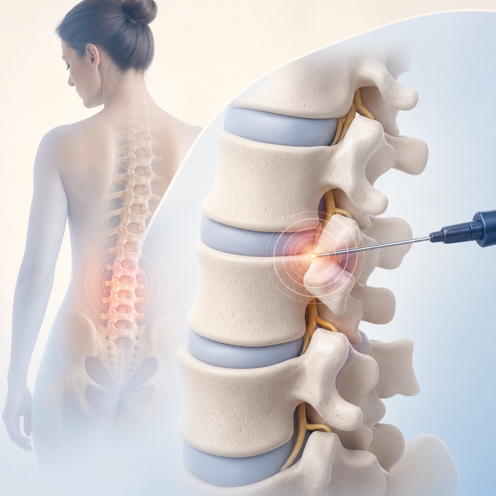
Nerve ablation
Radiofrequency Ablation for Back, Neck and Knee Pain
Radiofrequency ablation in Houston for facet (back, neck), genicular (knee) and SI joint pain: a heat lesion giving 6–12+ months of relief, repeatable.
In short
Radiofrequency ablation (RFA) uses a needle-tip electrode heated to about 80 °C for about 90 seconds to create a small heat lesion on a sensory nerve, interrupting the pain signal it carries. It is a treatment, done only after diagnostic nerve blocks have proven that the target nerve is the pain source. Relief typically lasts 6–12 months or longer, and because the nerve slowly regrows, the procedure can be repeated when pain returns. We use it for three targets: the medial branch nerves of the facet joints in the low back and neck, the genicular nerves of the knee, and the lateral branch nerves of the sacroiliac joint.
Key facts
| Treats | Facet joint pain in the low back or neck, knee pain from osteoarthritis or after knee replacement, and sacroiliac joint pain, each confirmed by diagnostic blocks |
|---|---|
| Test or treatment? | Treatment. The tests come first: medial branch blocks, genicular nerve blocks or SI lateral branch blocks |
| Procedure time | 30–60 minutes depending on the number of nerves; about 90 minutes at the office |
| Anesthesia | Local anesthetic plus light IV sedation for most patients; you are awake enough to answer during nerve testing |
| Downtime | 1–3 days of soreness; desk work within 1–2 days, physical work within about a week |
| When relief starts | Typically 2–4 weeks, after the treated area settles; some people notice it sooner |
| How long relief lasts | Typically 6–12 months, sometimes 18–24 months; the nerve regrows and pain gradually returns |
| How often it can be repeated | When pain returns, usually no sooner than 6 months after the last ablation; repeat ablations work about as well as the first |
| Insurance | Covered by Medicare and most plans after two positive diagnostic blocks (facet), one or two positive blocks (knee, SI); Medicare limits facet RFA to twice per year per spine region |
Who it is for
- No diagnostic blocks, or blocks with weak or inconsistent relief: ablation without a positive test rarely works and insurers will not authorize it
- Leg or arm nerve pain from a disc or stenosis; ablation treats joint pain, not radiculopathy
- Active infection or fever, or a skin infection over the treatment site
- Blood thinners that cannot be managed, or a bleeding disorder
- Pregnancy, because fluoroscopy uses X-ray
- An implanted pacemaker, defibrillator or spinal cord stimulator is not a bar, but it requires planning with your device team before the procedure
At a glance
| Procedure | Target nerves | Used for | Required test first | Typical relief |
|---|---|---|---|---|
| Lumbar facet RFA | Medial branches of the lumbar dorsal rami (L1–L5) and L5 dorsal ramus | Low back pain from facet joints, worse with extension and twisting | Two medial branch blocks with ≥80% relief | 6–12+ months; repeatable |
| Cervical facet RFA | Cervical medial branches (C3–C7), third occipital nerve for C2–3 | Neck pain from facet joints, whiplash, cervicogenic headache | Two medial branch blocks with ≥80% relief | 6–12+ months; repeatable |
| Genicular RFA | Superomedial, superolateral and inferomedial genicular nerves | Knee osteoarthritis not ready for replacement; persistent pain after knee replacement | Genicular nerve block with ≥50% relief | 6–12 months; repeatable |
| SI lateral-branch RFA | S1–S3 lateral branches and L5 dorsal ramus | Sacroiliac joint pain confirmed by exam and blocks | SI joint injection or lateral branch block with ≥50–75% relief | 6–12 months; repeatable |
How radiofrequency ablation works
Small sensory nerves carry pain from an arthritic facet joint, knee or SI joint to the spinal cord. If a diagnostic block has shown that numbing those nerves stops the pain, interrupting them for longer will stop it for longer. That is all RFA does. It is a heat lesion, not a "modulation": radiofrequency current passes through the exposed tip of a special needle, heats the surrounding tissue to about 80 °C, and holds it there for 60–90 seconds, coagulating a few millimeters of the nerve. The nerve is not removed; its outer sheath survives, and over 6–18 months the fibers regrow along it. That is why relief is long but not permanent, and why the procedure can be repeated.
Before any lesion is made, we test the needle position electrically. A sensory test (50 Hz) should reproduce your usual pain or a tingling in the right area; a motor test (2 Hz) confirms the needle is not next to a nerve that moves a leg or arm. Only then is the lesion made, which is why sedation stays light enough for you to answer.
Cooled radiofrequency circulates water through the needle so the tip can deliver more energy without charring, producing a lesion several times larger; it is used at the knee and SI joint, where the nerve's exact position varies. Conventional thermal RFA makes a smaller, precise lesion and is standard for medial branch nerves, whose location is predictable. Radiofrequency ablation is performed at our Houston and Webster offices, and in Pearland when that office opens in November 2026.
Lumbar and cervical facet (medial branch) RFA
This is the most common ablation we perform. Each facet joint is supplied by two medial branch nerves, so treating two joints on one side means three lesions; two-sided treatment doubles that. The target is the same nerve numbed during your medial branch blocks, which is why those blocks must be positive first.
Low back. Typical candidates have facet joint pain: back pain worse with arching, twisting or standing, without true sciatica. In the best-selected patients, about 6 in 10 have at least 90% relief a year later, and most have at least 50% relief for 6–12 months. Medicare allows facet RFA no more than twice per year per spine region, with at least 6 months between treatments of the same joints.
Neck. Cervical medial branch RFA follows the same logic for neck pain from the facet joints, including after whiplash. The landmark trial in whiplash patients found a median of about 9 months of relief after true ablation versus about a week after a sham procedure. Cervical RFA also treats cervicogenic headache arising from the upper cervical joints. After cervical ablation, a numb patch of skin on the back of the neck and a few weeks of neck-muscle soreness are common.
Genicular RFA for knee pain
The knee's sensory supply comes from several genicular nerves; three of them (superomedial, superolateral and inferomedial) sit at predictable spots on the femur and tibia and can be ablated without affecting the nerves that move the foot. A genicular nerve block with at least 50% relief comes first. Candidates are people with knee pain from moderate-to-severe osteoarthritis who are not ready for, or are not candidates for, knee replacement (age, weight, medical conditions, or preference), and people with persistent pain 6 or more months after a knee replacement once their surgeon has ruled out infection, loosening or instability.
In a randomized trial of cooled genicular RFA versus steroid injection, about 3 in 4 patients had at least 50% relief at 6 months after ablation compared with about 1 in 6 after steroid; relief typically lasts 6–12 months and the procedure can be repeated. Genicular RFA does not treat the arthritis itself, does not interfere with a later knee replacement, and has no effect on the joint's cartilage.
SI lateral-branch RFA for sacroiliac pain
The sacroiliac joint is supplied at the back by small lateral branch nerves from the S1–S3 sacral foramina and the L5 dorsal ramus. Their positions vary, so this ablation uses cooled radiofrequency or a strip-lesion technique to cover the area where the nerves run. Candidates have SI joint pain confirmed by provocation tests and at least 50–75% relief from an SI joint injection or lateral branch block.
In randomized sham-controlled trials of cooled SI RFA, roughly half of treated patients had at least 50% relief at 3–6 months versus about 1 in 8 with sham, and relief in responders typically lasts 6–12 months. Because the ablation treats only the back of the joint, it helps less when pain comes from the front of the joint or the ligaments; when SI pain returns repeatedly after good ablation results, SI joint fusion is the surgical option.
What happens on procedure day
- Check-in: we confirm your blood-thinner plan, allergies, any implanted device (pacemaker, defibrillator, stimulator), and your driver. A grounding pad is placed on your thigh or back; remove metal jewelry and tell us about any metal implants near the site.
- Positioning and sedation: face down for spine and SI ablations, on your back for the knee. An IV is placed and light sedation given if planned; you remain able to answer questions.
- Numbing: lidocaine in the skin and along each needle path. Ablation needles are slightly larger than block needles, so the numbing is thorough.
- Needle placement and testing: under live X-ray each electrode is placed at its target. Sensory stimulation should reproduce your usual pain or tingling in the right area; motor stimulation confirms no muscle twitching in the leg or arm. Positions are adjusted until both tests are right.
- Lesioning: local anesthetic is injected through the needle, then the tip is heated to about 80 °C for 60–90 seconds (cooled RF: a lower tip temperature for about 150 seconds). You may feel deep warmth or pressure; sharp pain means we pause and add anesthetic. Each nerve takes a few minutes.
- Recovery: 20–30 minutes of observation, checking leg or arm strength, then home with your driver. Plan on a quiet rest of the day.
After the procedure
Day 0. Rest, ice 15–20 minutes at a time, and use your usual pain medication. Numbness or heaviness near the site for a few hours is normal. No driving after sedation.
Days 1–7. Expect the treated area to feel sore, bruised or sunburned. A burning skin sensitivity over the site (post-procedure neuritis) affects roughly 1 in 10 people and lasts 1–2 weeks; it is a healing nerve, not a complication. Ice, an anti-inflammatory if you can take one, and gentle activity help. Most people are back to desk work in 1–2 days.
Weeks 2–4. The benefit becomes clear as the treated nerves stop transmitting. Start or return to physical therapy now; the pain-free window is the time to rebuild strength.
Judging the response. We check in at 4–6 weeks. Relief of 50% or more is a good result; little change by 6 weeks means the ablation missed or the diagnosis needs revisiting, and we discuss options rather than repeating it.
Call us for fever over 100.4 °F, redness, swelling or drainage at the sites, leg or arm weakness that persists beyond the day, or burning pain that is getting worse after two weeks.
What the evidence shows
Radiofrequency ablation has strong evidence when patients are selected with rigorous diagnostic blocks and mixed evidence when they are not; both facts matter.
- Facet RFA. A sham-controlled trial of cervical medial branch RFA (Lord and colleagues, NEJM 1996) produced a median 263 days of relief versus 8 days for sham. In carefully selected lumbar patients (Dreyfuss 2000), 6 in 10 had at least 90% relief at one year. ASIPP rates the evidence as Level II (moderate).
- The MINT trials (JAMA 2017) found no clinically important benefit from adding RFA to exercise at 3 months. Patients were selected with a single block at a 50% threshold, and the technique has been widely criticized. Our reading, shared with the 2020 multispecialty consensus guidelines, is that MINT shows what loose selection produces, which is why we require two blocks with 80% relief.
- Genicular RFA. In a multicenter randomized trial (Davis 2018), 74% of patients had at least 50% relief at 6 months after cooled genicular RFA versus 16% after steroid injection.
- SI lateral branch RFA. Two sham-controlled trials (Cohen 2008, Patel 2012) found roughly half of patients with at least 50% relief at 3–6 months versus about 1 in 8 with sham.
When pain returns after a good result, a repeat ablation succeeds in about 8 in 10 patients and lasts about as long as the first.
Alternatives and what comes next
Every ablation begins with the matching diagnostic block: medial branch blocks for the spine, a genicular nerve block for the knee, or an SI joint injection. Patients who prefer a single therapeutic injection can choose facet joint injections or knee injections, with shorter relief. If ablation works and wears off, it can be repeated. If SI pain keeps returning despite good ablation results, SI fusion is the surgical next step; for knee arthritis, knee replacement remains definitive, and genicular RFA does not interfere with it. Other nerve targets are on the nerve blocks hub.
Safety and preparation
- Blood thinners: facet, genicular and SI ablations are low-to-intermediate bleeding-risk procedures. Many patients can continue aspirin, and some other agents are continued or briefly paused case by case. Tell us about warfarin, apixaban, rivaroxaban, dabigatran, clopidogrel, ticagrelor and enoxaparin; we decide with your prescriber. Never stop a blood thinner on your own.
- Pacemaker, defibrillator (ICD) or implanted stimulator: tell us at scheduling. Radiofrequency current can interfere with cardiac devices, so we obtain guidance from your cardiology or device clinic, may use a magnet or have the device reprogrammed for the procedure, and place the grounding pad away from the device. Spinal cord or peripheral nerve stimulators are usually switched off during the ablation.
- Grounding pad and metal: the pad is placed on clean, dry, hair-free skin. Remove jewelry and body piercings near the site, and tell us about metal implants (hip or knee replacement, spinal hardware) so the pad and current path avoid them.
- Diabetes: no steroid is used in a standard ablation (a small dose is sometimes injected afterward to reduce neuritis; tell us if you prefer to avoid it). Eat normally unless sedation is planned.
- Infection or fever: we reschedule if you have a fever, active infection, or a skin problem over the treatment site.
- Allergies (contrast, steroid, local anesthetic): tell us about reactions to lidocaine, bupivacaine, X-ray dye or cortisone products.
- Pregnancy: the procedure uses X-ray; tell us if you are or could be pregnant.
- Sedation and driving: most patients receive light IV sedation. Nothing to eat for 6 hours or drink for 2 hours beforehand, and an adult driver is required. Even without sedation, plan for a ride; the treated area is sore afterward.
- Diagnostic blocks first: bring or confirm the pain diaries from your blocks; insurance authorization for ablation depends on them.
Risks and side effects
- Soreness, bruising or a sunburn-like burning over the treated area for 1–2 weeks (post-procedure neuritis), affecting roughly 1 in 10 people
- Temporary increase in pain for a few days before relief begins
- A numb or oddly sensitive patch of skin near the site, especially after cervical or knee ablation, lasting weeks to months
- Muscle soreness or a feeling of weakness in the back muscles for a few weeks (the medial branch supplies a small back muscle)
- Grogginess from sedation on the day
- Skin burn at the grounding pad or along the needle if the pad contact is poor; prevented by proper pad placement and no metal on the skin
- Bleeding or a small hematoma at the site
- Allergic reaction to local anesthetic, or to contrast if used
- Prolonged neuritis or burning pain lasting more than a month, usually settling with time or a short course of nerve-pain medication
- No benefit, or relief shorter than expected, most often when the diagnostic blocks were borderline
- Motor nerve injury, for example foot drop from lesioning near a spinal nerve root or the common peroneal nerve at the knee; prevented by motor stimulation testing before every lesion and by avoiding the inferolateral genicular nerve
- Infection at the site or deeper
- Deafferentation pain: a new, persistent burning pain from the treated nerve
- Interference with a pacemaker or defibrillator during lesioning, or heating of a nearby implanted lead; prevented by device planning, bipolar technique or magnet use as advised by your cardiologist or device manufacturer
- Charcot-like accelerated joint damage at the knee has been raised as a theoretical concern after genicular ablation; it has not been shown in trials, and we monitor for it
- Spinal cord or nerve root injury with cervical ablation, avoided by imaging in two planes and stimulation testing
Frequently asked questions
How long does radiofrequency ablation last?
Typically 6–12 months, and sometimes 18–24 months. The treated nerve regrows slowly along its sheath, and pain gradually returns as it does. When that happens the ablation can be repeated, and repeat treatments work about as well as the first.
Is radiofrequency ablation painful?
The procedure itself is done with local anesthetic and light sedation; most people feel pressure and deep warmth rather than sharp pain, and we add anesthetic if anything is sharp. Afterward, expect 1–2 weeks of soreness or a sunburn-like burning over the area while the treated nerve settles. That phase is normal and passes.
How soon does RFA work?
Some relief can be immediate from the local anesthetic, but the real benefit usually appears at 2–4 weeks, after the post-procedure soreness fades. We judge the result at 4–6 weeks.
Do I have to have medial branch blocks before ablation?
Yes. Ablation only works if the treated nerves are the ones carrying your pain, and the blocks are the only way to prove that. Medicare and most insurers require two positive blocks for facet ablation, and at least one positive block for knee and SI ablation, before they will authorize the procedure.
Can I have RFA if I have a pacemaker?
Usually yes, with planning. We contact your cardiology or device clinic for instructions, which may include reprogramming or a magnet during the procedure, and we position the grounding pad so the current path stays away from the device. Tell us about any implanted device when you schedule.
What is the difference between radiofrequency ablation and a nerve block?
A nerve block uses local anesthetic to numb a nerve for hours and is a test. Ablation uses heat to interrupt the same nerve for months and is the treatment that follows a positive test.
Does nerve ablation cause permanent damage or numbness?
The lesion is small and the nerve regrows over 6–18 months. A patch of skin numbness near the site is common for a few weeks to months, especially in the neck and knee. Permanent damage to nerves that control movement is rare because every needle is tested with motor stimulation before a lesion is made.
Can I have genicular RFA if I might need a knee replacement later?
Yes. Genicular ablation treats only the sensory nerves outside the joint, does not affect the bone or cartilage, and does not interfere with a future knee replacement. It is also used for pain that persists after a replacement once the surgeon has excluded mechanical causes.
Sources
- Percutaneous Radio-Frequency Neurotomy for Chronic Cervical Zygapophyseal-Joint Pain (Lord SM, et al., 1996) (opens in new tab) — New England Journal of Medicine
- Effect of Radiofrequency Denervation on Pain Intensity Among Patients With Chronic Low Back Pain: The MINT Randomized Clinical Trials (2017) (opens in new tab) — JAMA
- Consensus practice guidelines on interventions for lumbar facet joint pain from a multispecialty, international working group (2020) (opens in new tab) — Regional Anesthesia & Pain Medicine (Cohen SP, et al.)
- Prospective, Multicenter, Randomized, Crossover Clinical Trial Comparing the Safety and Effectiveness of Cooled Radiofrequency Ablation With Corticosteroid Injection in the Management of Knee Pain From Osteoarthritis (Davis T, et al., 2018) (opens in new tab) — Regional Anesthesia & Pain Medicine
- Randomized placebo-controlled study evaluating lateral branch radiofrequency denervation for sacroiliac joint pain (Cohen SP, et al., 2008) (opens in new tab) — Anesthesiology
Next step
Request an appointment
Call (832) 916-2075 or request an appointment online. New patients are welcome; the team confirms insurance and referral requirements before scheduling.