
Spine and bone health
Spinal Compression Fractures (Vertebral Compression Fractures)
Sudden back pain after a minor strain may be a spinal compression fracture. Houston pain specialists on the kyphoplasty window, red flags and bone care.
A vertebral compression fracture is a collapse of the front part of one of the bones of the spine. In older adults the cause is almost always osteoporosis, and the trigger can be as small as a cough, lifting a grocery bag, or a slip that did not even cause a fall. About 700,000 happen in the United States each year. Most heal with bracing, pain control and time, but a fracture that stays painful past a few weeks can be stabilized with kyphoplasty, a needle procedure that works best inside a window of roughly 6 to 8 weeks. Every fracture also means the bone itself needs treatment so the next one does not happen.
Key facts
| What it is | A crack and collapse of a vertebral body, most often in the mid-back (T11 to L2). The bone loses height in front, producing a wedge shape and a forward stoop. |
|---|---|
| Most common causes | Osteoporosis (over 85 percent), then trauma in younger people, long-term steroid use, and, in a minority, cancer or multiple myeloma weakening the bone. |
| Typical course | Acute pain is severe for 2 to 4 weeks and usually improves substantially by 6 to 12 weeks as the bone heals. About one-third of fractures remain painful longer. |
| See a specialist when | Pain is still severe at 2 to 3 weeks despite bracing and medication, you cannot get out of bed or walk, or imaging shows a fracture that is still unhealed (edema on MRI). |
| Treatments we offer | Pain control and bracing coordination, kyphoplasty and vertebroplasty for fractures that stay painful, and coordination of osteoporosis evaluation and treatment with your primary care physician or endocrinologist. |
When to get emergency care
Go to an emergency room or call 911 if you have:
- New weakness in the legs, trouble walking, numbness below the fracture, or loss of bladder or bowel control (fracture fragments may be pressing on the spinal cord or nerves)
- Back pain with a history of cancer, unexplained weight loss, night pain that does not ease lying still, or a fracture above the mid-back (T5 or higher) without a fall (possible pathologic fracture from tumor or myeloma, which needs a different workup)
- Fever with new back pain, especially with diabetes, recent infection or IV drug use (possible spinal infection)
- Severe pain after a high-energy injury such as a car accident or fall from height (possible unstable burst fracture)
- Pain so severe you cannot stand, eat or drink normally, particularly if you live alone
Most osteoporotic compression fractures are stable and do not threaten the spinal cord. The list above is the small group that needs emergency imaging first.
What a compression fracture is
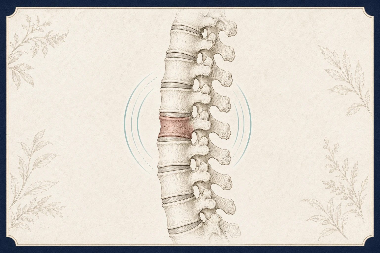
Each vertebra has a block-shaped front part, the vertebral body, that carries most of your weight. When bone becomes porous from osteoporosis, that block can crush under a load it once handled easily. The front collapses more than the back, so the bone becomes a wedge and the spine tips forward a little at that level. Several of these fractures over the years produce the rounded upper back (kyphosis) and height loss common in older adults.
These fractures are common: an estimated 700,000 occur in the United States each year, and about a quarter of women over 70 have at least one. Many are silent and are found on a chest X-ray done for another reason. Having one fracture raises the chance of another vertebral fracture in the next year to roughly one in five, which is why bone treatment matters as much as pain treatment.
A fracture is called pathologic when the bone broke because a tumor, multiple myeloma or infection weakened it rather than osteoporosis. These are a minority but change everything about treatment, so we look for them specifically.
Common causes
- Osteoporosis. The cause in more than 85 percent of cases. Risk rises with age, menopause, low body weight, smoking, heavy alcohol use, a parent who broke a hip, and prior fractures of any kind.
- Long-term steroid use. Prednisone and similar medications, used for asthma, rheumatoid arthritis, lupus, transplants and other conditions, thin bone quickly. Even 5 mg a day for 3 months raises fracture risk.
- Other medical causes of weak bone. Hyperparathyroidism, low vitamin D, celiac disease, chronic kidney disease, and some cancer and seizure medications.
- Trauma. In younger people with normal bone it takes a car accident, a fall from height or a sports injury to compress a vertebra.
- Cancer and myeloma. Breast, lung, prostate, kidney and thyroid cancers spread to the spine, and multiple myeloma grows inside bone. A fracture can be the first sign of either.
Symptoms and how it differs from look-alikes
The classic story is sudden, sharp mid- or low-back pain after a small effort in someone over 60, worst when standing or walking and better lying flat. The spot over the fracture is tender to a firm tap. Pain often wraps around the ribs or flank on one or both sides, which sometimes sends people to the emergency room for a kidney or heart problem first.
- Versus a muscle strain: strains hurt with movement but rarely make it impossible to stand, and are not tender to percussion over a single bone.
- Versus a herniated disc: disc pain usually radiates into a leg and is worse sitting; compression fractures rarely cause leg symptoms unless a fragment is pushed backward.
- Versus facet joint pain: facet pain is a chronic ache worse with leaning back, without the sudden onset.
- Versus a pathologic fracture: cancer-related pain is more constant, often worse at night, and may come with weight loss, fatigue or anemia. Fractures above T5 or in someone under 50 without trauma also raise concern.
How we diagnose it
X-rays show the loss of height and are often all that is needed to confirm a fracture. Comparing with older films tells us whether the fracture is new.
MRI with STIR sequences is the key test for deciding on treatment. Fresh, unhealed bone shows bright fluid signal (edema); a healed fracture does not. Edema is what tells us kyphoplasty can still help. MRI also shows whether any bone has been pushed backward toward the spinal cord and helps distinguish an osteoporotic fracture from a tumor. If you cannot have an MRI, a bone scan or CT is used instead.
Pathologic fracture workup. If the history, fracture pattern or MRI raises concern for cancer, we order blood work (complete blood count, calcium, serum and urine protein electrophoresis for myeloma, PSA in men) and may recommend a biopsy, which can be done through the same needle at the time of kyphoplasty. Findings go to your oncologist or primary care physician promptly.
Bone density (DEXA) and labs. A compression fracture with minimal trauma is itself enough to diagnose osteoporosis regardless of the DEXA score, but the scan sets a baseline and vitamin D, calcium, kidney and thyroid tests uncover treatable causes.
Neurologic exam of strength, sensation and reflexes in both legs at every visit, because a fracture can settle further in the first weeks.
Treatment options, in order
Treatment has two tracks that run at the same time: settling the pain of this fracture, and strengthening the bone to prevent the next one.
- Conservative care (first 2 to 4 weeks)
Short, scheduled pain medication (acetaminophen, a limited course of an anti-inflammatory if your kidneys and stomach allow, and short-term opioids only if needed to get out of bed and walk), a brace for comfort during the acute phase, and early walking. Prolonged bed rest speeds bone loss and raises the risk of clots and pneumonia. Calcitonin nasal spray is an option for acute fracture pain in the first weeks.
- Kyphoplasty or vertebroplasty (typically within 6 to 8 weeks)
If pain is still severe at 2 to 3 weeks despite the above, MRI shows edema and the pain matches the fracture level, a needle is guided into the vertebra under X-ray and bone cement is injected to stabilize it. In kyphoplasty a balloon first creates a cavity and restores some height. Most patients notice major pain relief within 24 to 48 hours. Randomized trials in acute, edema-positive fractures show clear benefit over sham or conservative care; trials that included older, partly healed fractures showed little difference, which is why timing matters. This is a treatment, not a test. Learn more →
- Osteoporosis treatment (starts now, continues for years)
Every patient with a low-trauma compression fracture qualifies for prescription bone treatment. Options include bisphosphonates (alendronate, zoledronic acid), denosumab, and for severe cases bone-building agents such as teriparatide, abaloparatide or romosozumab, along with vitamin D and calcium. These cut the risk of another vertebral fracture by roughly 50 to 70 percent. We coordinate this with your primary care physician, endocrinologist or a fracture liaison program rather than leaving it to a later visit.
- Physical therapy and fall prevention
Once acute pain settles, extension-based strengthening, balance training and a home safety review reduce both future fractures and the stooped posture that follows several fractures. See how we coordinate physical therapy. Learn more →
- Surgical referral
Reserved for unstable fractures, neurologic compromise, or progressive collapse and deformity. These are uncommon in osteoporotic fractures and are managed with a spine surgeon.
What you can do now
- Walk short distances several times a day, even during the painful phase; lying still for days weakens bone and muscle further.
- Log-roll to get out of bed: bend your knees, roll to your side as one unit, then push up with your arms.
- Avoid bending forward at the waist and lifting anything heavier than a gallon of milk until cleared.
- Take 1,000 to 1,200 mg of calcium daily from food and supplements combined and 800 to 1,000 IU of vitamin D unless told otherwise.
- Ask your primary care physician for a DEXA scan and a fracture-risk (FRAX) assessment if you have not had one; bring the report to your visit.
- Bring your X-rays and MRI on disc; the MRI timing determines your options.
We evaluate spinal compression fractures at our Houston and Webster offices, and in Pearland after that office opens in November 2026.
Frequently asked questions
How do I know if my back pain is a compression fracture?
Sudden sharp pain in the mid- or low-back after a minor strain, in someone over 60 or on long-term steroids, that is worse standing and better lying down, with tenderness when the spine is tapped, is the typical picture. An X-ray confirms it; an MRI tells us how fresh it is.
Do compression fractures heal on their own?
Most do. The bone knits over 6 to 12 weeks and pain improves along with it, though the lost height does not come back. About a third of people have pain that persists beyond that, and those are the patients who benefit from kyphoplasty.
Why is the timing for kyphoplasty important?
Cement stabilizes a fracture that is still moving. Once the bone has healed, usually by 8 to 12 weeks, there is little left to stabilize and the trials show little benefit. MRI edema is how we know the fracture is still active. The best results come from treatment within about 6 to 8 weeks of the fracture, and sooner if pain is disabling.
Does having a compression fracture mean I have cancer?
Usually not. More than 85 percent are from osteoporosis. We look harder for cancer or myeloma when there is a history of cancer, weight loss, night pain, a fracture high in the mid-back, a patient under 50 with no injury, or MRI features that do not fit osteoporosis. When needed, a biopsy can be taken during kyphoplasty.
Should I be treated for osteoporosis even if my bone density test looks okay?
Yes. A vertebral fracture from a minor strain is a diagnosis of osteoporosis on its own, whatever the DEXA score says. Medication after a fracture cuts the risk of another vertebral fracture by half or more, and the highest-risk period is the first year.
Will I get shorter or develop a hump?
One fracture usually costs a centimeter or two of height. Multiple fractures produce the rounded upper back called kyphosis, which also reduces lung capacity and balance. Treating the bone and strengthening the back extensors is what prevents that progression.
Is kyphoplasty covered by insurance?
Medicare and most commercial plans cover kyphoplasty and vertebroplasty for painful osteoporotic or pathologic compression fractures when conservative care has failed and imaging shows an active fracture, usually with documentation of pain severity and functional limitation. We handle the authorization.
Sources
- Osteoporosis and Spinal Fractures (opens in new tab) — American Academy of Orthopaedic Surgeons, OrthoInfo
- Osteoporosis: overview, diagnosis and treatment (opens in new tab) — National Institute of Arthritis and Musculoskeletal and Skin Diseases (NIAMS)
- Vertebroplasty versus conservative treatment in acute osteoporotic vertebral compression fractures (VERTOS II, Klazen CA et al., 2010) (opens in new tab) — The Lancet / PubMed
- Efficacy and safety of balloon kyphoplasty compared with non-surgical care for vertebral compression fracture (FREE trial, Wardlaw D et al., 2009) (opens in new tab) — The Lancet / PubMed
- Percutaneous vertebroplasty for osteoporotic vertebral compression fracture (Cochrane review, 2018) (opens in new tab) — Cochrane Database of Systematic Reviews
Request an appointment
Call (832) 916-2075 or request an appointment online. New patients are welcome; the team confirms insurance and referral requirements before scheduling.